
Sternal cleft, or cleft sternum, is a rare congenital malformation of the chest wall. This condition is characterized by a complete or partial splitting of the sternum. Understanding the causes of this malformation and its structural effects is crucial for better treatment and surgical planning.
Causes of Sternal Cleft
Cleft sternum occurs during embryogenesis, and its precise etiology is not fully understood. The sternum usually forms when two lateral mesenchymal bars come together and move toward the midline during early fetal development. A sternal cleft may form if any part of this fusion process is not normal. Once a sternal cleft forms, it is a permanent deformity that cannot heal or fuse on its own after birth.
Classification and Physical Presentation
There are two kinds of sternal clefts: complete and partial. And two types of partial clefts exist: superior and inferior. Superior sternal cleft is more common and usually has a U or V shape. Inferior sternal cleft is often linked to intricate syndromes, including Cantrell’s Pentalogy (Pentalogy of Cantrell).
Symptomatology depends on defect type and associated anomalies. Isolated sternal cleft is rare and may manifest as tachypnea or mild exercise intolerance. Most cases are complex with significant symptoms. Limited partial sternal clefts may show only visible chest pulsations; extensive defects cause paradoxical breathing and recurrent respiratory infections. Inferior sternal cleft is typically present with Cantrell’s Pentalogy, whose greatest hazard lies in various types of cardiac malformations, especially ventricular septal defect. Complete sternal cleft exhibits prominent cardiac pulsations and severe cardiopulmonary compromise, particularly when complicated by intrinsic cardiac defects.
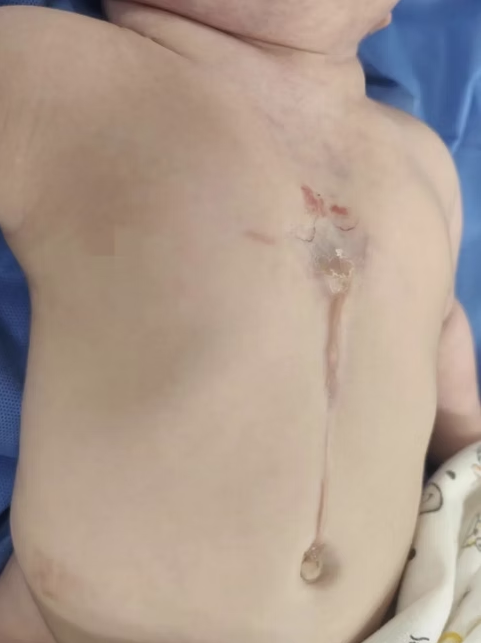
Surgical Correction for Special Needs
The Institute of Chest Wall Surgery (ICWS) provides specialized diagnosis and surgical treatment for sternal clefts.
For isolated sternal cleft without associated malformations and with a narrow gap between the residual bony edges, direct repair may be considered. In more complex cases, advanced reconstructive techniques involving autologous rib grafts or synthetic materials are used to repair the defect and restore thoracic integrity. When cardiac anomalies are present, surgical repair must be carefully coordinated, either simultaneously or in a staged manner, to ensure optimal outcomes while avoiding complications such as cardiac compression.